The human urinary system, with its multiple nerve endings, is a complex mechanism. An important element in its work is the occurrence of the urge to withdraw urine, the ability of a person to control the restraint and relaxation of muscles. This process is guaranteed by the innervation of the bladder (in other words: its connection with the central nervous system). Special impulses are transmitted through nerve tissues, giving peculiar signals about its filling.
The bladder is a hollow organ located in the pelvis. It serves as a kind of reservoir for the accumulation of urine (urine) produced by the kidneys for the purpose of its further removal from the body.
Thanks to innervation, a person can restrain emptying the bladder for a certain time by force of will. Disruption of the nervous system leads to a malfunction in the well-established urinary system, which can lead to neurogenic syndrome.
Urinary excretion algorithm
To better understand the topic raised, let's look at the detailed mechanism of urination. The average urea volume of an adult is 500 ml. However, the volume indicator may vary in both sexes. In males it reaches 750 ml, and in females it does not exceed 550 ml.
So, the continuous functioning of the kidneys is carried out from time to time by filling the urea with urine. Its ability to expand the walls allows urine to fill the bladder up to 150 ml without causing any discomfort. When the liquid exceeds the specified volume, the organ cavity enters the stretching stage, and the pressure in it increases and the desire to urinate arises.
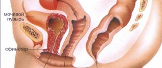
The reaction to a stimulus occurs at the reflex level. In the adjacent area of the urethral canal and bladder there is an internal sphincter, and just below the external one. When the organ cavity is not filled with excess fluid and is not under pressure, the muscles of the internal and external sphincters are closed, thereby preventing the spontaneous release of urine. When signals for emptying appear, the valves weaken, the walls of the bladder contract, and urine is released.
Diseases provoked by the innervation of an organ in a state filled and empty of urine
Excess of innervation leads to a neurogenic bladder. This disease indicates the beginning of incorrect functioning of the urinary canals. Urinary tract problems can be acquired during life or can be a congenital disorder related to the nerves.
The connection between the bladder and the nervous system is very important for a person to live a full life. When the disease occurs, the patient’s urinary canals atrophy, or they work too actively. Such disorders can manifest themselves with injuries or parallel diseases (pathologies of the anterior part of the central nervous system, multiple sclerosis, stroke, parkinsonism, Alzheimer's disease, spinal cord lesions). The patient completely loses control over the process of removing urine from the body.
In turn, the neurogenicity of the muscular organ is divided into hyperactive and hypoactive types of disease development.
Disease model
The interaction of the urinary organ with the central nervous system occurs due to the presence of several types of nerves in it:
- sympathetic;
- parasympathetic;
- sensory fibers.
The walls of the bladder are equipped with many receptor nerve pathways, scattered neurons of the ANS (autonomic nervous system), as well as nerve ganglia. The performance of the listed elements is the basis for the regular management of urine output, since each of them performs a specific task. Various disorders of the innervation of the bladder can lead to a variety of disorders.
Parasympathetic innervation
The parasympathetic center (excitatory fibers) is located in the sacral part of the spinal cord. This is where the prenodal fibers (preganglionic) begin, which form the pelvic plexus and participate in the innervation of the pelvic organs.
The fibers activate the nerve nodes (ganglia), which are located in the walls of the bladder, then the smooth muscle contracts, as a result of which the sphincter muscles are weakened, and urine is released.
Sympathetic type of disease
The cells of the autonomic nervous system, which take part in the secretion of fluid, are located in the intermediate lateral column of the lumbar region of the spinal cord. The main task is to stimulate the closure of the neck, due to which fluid collects in the cavity of the bladder.
Therefore, sympathetic (retaining) nerve endings are found in significant numbers in the triangle of the urinary system organ and the cervix. Sympathetic fibers have almost no effect on the process of urine excretion.
The role of sensory nerves
The pelvic nerves include sensory nerve fibers that send signals about how much the walls of the bladder are stretched. The strongest of them come from the posterior part of the urethral canal. Actually, they are responsible for the appearance of the reflex to empty the urinary cavity.
Paruria (urination) reflex
As the bladder fills, rapid fluctuations appear in the form of the reaction of myocytes to the influence of the electrochemical impulse. Stimulates reflex contractions by activating the stretching nerve endings of the posterior urethra. Nerve impulses from the receptors are carried to the sacral segments (roots) of the dorsal brain along the pelvic nerves.
The urination reflex is a set of periodically repeating processes.
- As the bladder fills with urine, the pressure increases.
- The contraction of the bubble sets in motion the stretching.
- The flow of pulsation increases and intensifies contractions of the bladder wall.
- Impulses from contractions are transferred along the pelvic nerves to the roots of the spinal cord, and the central nervous system forms the urge to paruria.
- Contractions of the bladder during urination relax the detrusor, and the pressure stabilizes.
The paruria reflex will increase until the act of emission of urine occurs.
Disturbance of the nervous regulation of urination
Failures in the innervation of an organ can manifest themselves in 3 cases:
- Hyperreflex bubble . Urine cannot accumulate even to a minimum volume and begins to be excreted immediately. Because of this, a person feels an increased urge to urinate, while the amount of urine emitted is minimal. This disorder is a consequence of serious problems with the central nervous system.
- Hyporeflexivity of the organ . This means that excess fluid accumulates (up to 1.5 liters), and emptying the bladder causes difficulty. Hyporeflexivity can provoke inflammation and the development of infection in the kidneys. This type of innervation disorder is characterized by damage to the sacral part of the brain.
- Areflexivity . In this case, a person cannot control the process of urination, since it occurs spontaneously at the moment of extreme accumulation of urine in the cavity.
All 3 cases are due to different reasons. The most common are:
- traumatic brain injury;
- heart diseases;
- vascular disease;
- neoplasms in the brain;
- multiple sclerosis.
Recognizing a pathological condition based only on external signs is problematic. The form of the disease is directly related to the area of the brain that has undergone abnormalities.
To indicate disruption of the activity of the urine storage tank due to nervous disorders, a special term was introduced in medical practice - “neurogenicity of the bladder.” Different types of nerve tract lesions have their own effect on the removal of urine from the human body, which will be discussed further.
Innervation of the bladder
The transmission of impulses is provided by the autonomic nervous system, dendrites and roots. The main connection between the bladder and the central nervous system is provided by somatic nerves connected to each other and forming Pelvic nerves consist of afferent (sensitive) and efferent (motor) fibers. Signals about the degree of stretching of the bladder are transmitted along afferent fibers. Impulses emanating from the posterior urethra contribute to the activation of reflexes oriented towards urination.
Emptying the bladder can be reflexive or voluntary. Unconditional urination is carried out thanks to neurons of the sympathetic and parasympathetic innervation. Centripetal units of nervous tissue are responsible for meaningful urination. When an organ is filled with urine, pressure increases, excited sensors send a signal to the dorsal brain, and then to the cerebral hemispheres.
Brain lesions that disrupt innervation
The following brain pathologies can disrupt innervation:
- Multiple sclerosis . The functioning of the lateral, posterior columns of the cervical spinal cord leads to failure. In 50% of patients, the fact of involuntary emission of urine was revealed. The main symptoms develop over time. Sequestration of an intervertebral hernia of the initial stage entails a suspension of urine output and difficulty in emptying the organ. Signs of irritation then appear.
- Supraspinal disorders of the motor systems of the brain . They specifically affect the reflex function of urination. Symptoms: enuresis, frequent urges, including at night. But, since the coordination of the functioning of the basic muscles of the urinary tract is preserved, the pressure inside it remains within normal limits, thereby excluding the development of urological diseases.
- Peripheral paralysis . Prevents reflex muscle contraction, which prevents the lower sphincter muscle from relaxing on its own.
- Diabetic neuropathy . It disrupts the functioning of pushing urine out of the organ cavity.
- Lumbar spinal stenosis . It affects the urinary system depending on the variety and level of the destructive process.
- Cauda equina syndrome . It can cause incontinence due to overcrowding of the bladder, as well as a suspension of urine output from the body.
- Hidden spinal dysraphism (SSD) . This entails a failure of bladder reflexion, so the person cannot control urination. The release of fluid begins spontaneously when the organ is overfilled with fluid.
Variants of dysfunction in cases of significant brain damage
Consequences for the urinary system with complete damage to the spinal cord:
- Disrupted activity of the suprasacral areas of the spinal cord caused by a neoplasm, inflammation, or injury. First, hyperreflexia of the muscular membrane of the organ (detrusor) develops, after which independent contraction of the sphincter and bladder muscles begins. As a result, the pressure in the organ increases, and the volume of fluid at the outlet is scanty.
- Damage to the activity of the sacral elements of the spinal cord due to injury, hernia. The frequency of urges decreases, and urine output becomes difficult. The process of urination becomes uncontrollable for a person. Spontaneous release of fluid occurs when the bladder is overcrowded.
The effect of innervation disturbances on the urinary tract
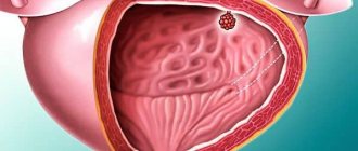
With improper innervation, the blood supply to the urinary tract organs is disrupted. Thus, with a neurogenic bladder, cystitis is often associated, which can cause microcysts.
Microcysts are a decrease in the size of the bladder due to chronic inflammation. With microcysts, bladder function is significantly impaired. Microcysts are one of the most complex complications of chronic cystitis and neurogenic bladder.
If urine remains in the bladder, the risk of inflammatory diseases of the urinary tract increases. If the neurogenic bladder is complicated by cystitis, then this poses a health hazard and sometimes requires surgical intervention.
Diagnostics
Diagnosis of the disease begins with a detailed interview with the patient. The following methods can be used:
- The patient is asked to keep a urination log for several days, in which the time and volume of fluid consumed will be noted.
- A thorough examination of the urinary system for the presence of infection by taking a urine sample.
- Examination of the internal organs of the system using: ultrasound, MRI, x-ray with preliminary administration of a contrast agent. Such methods are necessary to exclude symptoms of the inflammatory process and various anomalies of the urinary tract.
- Examination of the nervous system to establish (refute) the pathological condition of the brain and spinal cord using CT, MRI.
Cases cannot be excluded when the examination cannot find the cause, then the conclusion speaks of a neurogenic bladder of unknown origin.
Treatment
Treatment is determined by the nature of the lesion, its degree and happens:
- medicinal;
- surgical;
- non-medicinal.
Innervation is restored in full when using various treatment methods.
- Therapy to activate all parts of the nervous system using the following means:
- cholinomimetics;
- coenzymes;
- andrenomimetics;
- drugs: Aceclidine, Citrochrome C, Isoptin.
- A method of electrical stimulation of the urinary system in order to activate the work of the sphincters.
- The use of antidepressants and tranquilizers to support autonomic regulation.
Urgent hospitalization is required when urine output completely stops. In this case, catheters are used to remove it until it is completely cured. Innervation can be restored in full. During the treatment period, sleep patterns, walks, and gymnastics are important.
The urinary process is closely related to the state of the central nervous system. If nerve impulses are disrupted in one of its sections, innervation of the bladder may occur. It is important to see a doctor, timely diagnosis and therapy. Communication with the central nervous system can be restored in full.
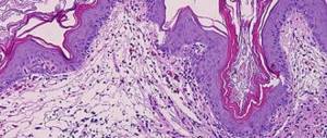
The trophic function of nerves is less important for the normal functioning of tissues than the blood supply, but at the same time, disruption of innervation can lead to the development of superficial necrosis - neurotrophic ulcers.
A feature of neurotrophic ulcers is a sharp inhibition of reparative processes. This is largely due to the fact that it is difficult to eliminate or at least reduce the influence of the etiological factor (impaired innervation).
Neurotrophic ulcers can form due to damage and diseases of the spinal cord (spinal injury, syringomyelia), damage to peripheral nerves.
Hyperactive appearance of the neurogenic organ for storing and excreting urine
This type of development of neurogenic bladder malaise entails impaired functioning of the part of the nervous system that is located above the bridge of the anterior part of the central nervous system. In this case, the tension in the muscles of the urinary system becomes more intense.
Doctors diagnose such phenomena as detrusor hyperreflexia. This type of excess of the innervation of the saccular organ leads to uncontrolled urination; involuntary release of urine can begin at any inopportune moment. The disease causes severe social and psychological discomfort to a person.
When a patient has an overactive detrusor disease, urine does not accumulate in the hollow organ, so he is forced to visit the restroom many times.
Diagnosis and timely therapy
The patient must undergo general blood and urine tests to identify a possible inflammatory process. You should also undergo a number of procedures for a comprehensive examination of the urinary tract, such as: ultrasound, urethrocytography, cytoscopy, urography, MRI if necessary and others.
To study neurological abnormalities, you may be prescribed an EEG, MRI, or examination using other methods. Neurogenic bladder disease is often cured. The main thing is to contact a specialist in time. As for medications, you may be prescribed medications that improve blood circulation, antibiotics, adrenergic blockers, and anticholinergics.
Hypoactive type of neurogenic muscle bladder
This type of disease begins its development under the pons of the brain, in most cases the lesion occurs in the sacral region. Such defectiveness of the nervous system results in incomplete contractions of the lower urinary excretory muscles or a complete absence of the necessary contractions. Doctors diagnose this course of the disease as detrusor areflexia.
Patients are simply not physiologically able to go to the toilet normally when the organ is full. They lose the sensitivity of the emptiness of the muscular organ, suffering from pain in the urethra. Some people do not feel the urge to urinate and cannot control the orbicularis muscle, which serves to narrow or close the urinary duct.