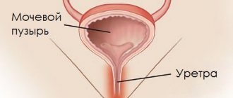
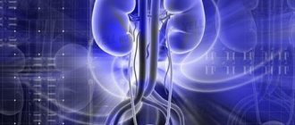
The bladder sphincter is directly involved in the process of formation and removal of urine from the body. The functioning of the genitourinary system and the general condition of a person depend on its condition and level of functionality. Pathologies of the organ lead to the appearance of an unpleasant problem - enuresis (urinary incontinence).
The bladder sphincter is a key part of the organ responsible for the outflow of urine.
Sex differences
Main articles: Male external urethral sphincter and Female external urethral sphincter
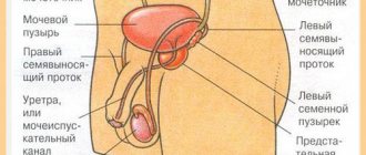
In males and females, both the internal and external urethral sphincters function to inhibit the release of urine. In men, the internal urethral sphincter functions to prevent reflux of seminal fluid into the male bladder during ejaculation.
Females have more complex external sphincter muscles than males as it consists of three parts: the sphincter urethrae, the urethrovaginal muscle, and the compressor urethrae. Urethrovaginal muscle fibers wrap around the vagina and urethra and contraction results in a narrowing of both the vagina and urethra. The origin of the compressor is the urethrae muscle on the right and left below the pubic ramus and wraps anteriorly around the urethra so when it contracts it compresses the urethra away from the vagina. The external urethrae, like in men, wraps exclusively around the urethra.
Congenital abnormalities of the female urethra can be surgically repaired with colpoplasty.
Sphincters located in the large intestine
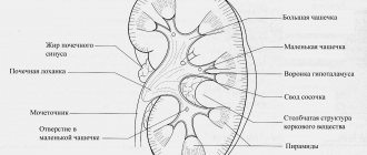
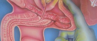
The normal functioning of the large intestine directly depends on the coordinated functioning of all the departments that make up its composition. The most important muscle structure is the sphincter. At the base of the human colon there are five main sections.
- Ascending colon.
- Descending colon.
- Transverse.
- Straight.
- Sigmoid.
Some areas of the large intestine are transitions between its sections. This is a kind of division, characterized by the presence of bundles consisting of smooth muscles. They work voluntarily and involuntarily.
The colon contains the following sphincters:
- ileocecal;
- Busie;
- Girsha;
- Cannon-Behm;
- Bally;
- sigmo-rectal;
- Rossi-Muthier.
The ileocecal sphincter muscles are located between the small and large intestines. Buzi is localized in the zone of transition of the cecum to the ascending colon. It can be examined during an x-ray examination. Hirsch is located at the upper part of the colon. Cannon-Behm is located at its beginning. Bali occupies the space between the sigmoid and descending colon.
The sigmo-rectal sphincter of the anus separates the rectum and sigmoid colon. Rossi-Muthier is represented by a circular tissue bundle consisting of smooth muscles.
Preparation for the procedure
Before the procedure, an ultrasound is prescribed, which allows you to determine the diameter of the organ, the location of the stone or the characteristics of the narrowing. In addition, you will need to undergo a general urine and blood test and undergo a computed tomography scan. Sometimes it is necessary to consult with other specialists, for example, a therapist or gynecologist for women. If there are no contraindications, a date for the examination is set. A few days before your visit to the doctor, avoid alcoholic beverages, and on the day of the procedure you should avoid food. If you take any medications on an ongoing basis, be sure to inform your specialist. Before inserting the instrument, the patient is dressed in a sterile uniform, after which he is placed in a reclining position on a special chair. The procedure is carried out by a doctor and his assistants.
Functional features of muscle structure
The anal sphincter is based on two main parts: external and internal. The outer section is represented by many receptors responsible for its stretching. This area is completely controlled by the human brain. In terms of its external characteristics, the structure resembles a ring up to 10 cm long. It is based on three layers of muscles, localized in the superficial and deep layers.
The inside of the anal sphincter is beyond the control of the human brain. Its contraction is carried out under the influence of physical force. This part is responsible for containing gases and liquids until a person makes an effort to eliminate them. This function is provided by sphincter fibers.
The muscle tone of the organ and its unique features allow you to completely control the act of defecation. This is ensured due to the presence of muscle bundles. Under the influence of certain forces, they contract, which allows the contents of the intestine to come out.
Exercises for the treatment of cystitis
In the complex therapy of inflammation of the bladder tissues with cystitis, an important element of treatment is gymnastic exercises. Properly selected exercises for cystitis can influence a variety of irritants in the body - pathogenic and physiological, involving mechanisms involved in pathological processes in the general process of reactivity. Therefore, exercise therapy is quite rightly considered one of the methods of pathogenetic treatment of cystitis.
The basis for the effectiveness of treatment of acute and chronic cystitis is the restoration of adequate blood supply to the bladder walls. The flow of blood to tissues affected by inflammation provides sufficient immune phagocytosis (protection), producing antibodies against infection and stimulating the adrenal glands to secrete special substances that promote restoration processes in the affected cells, clearing the inflammatory zone of dead and destroyed tissue cells by inflammation, improving metabolic processes.
Features of exercise therapy for cystitis
The importance of using exercise therapy for cystitis of its various forms has been confirmed by numerous studies by foreign and domestic scientists. The special effectiveness of therapeutic exercises when performed comprehensively and regularly is emphasized. To achieve positive results, the exercise therapy regimen must contain exercises that alternately affect all muscle groups in the body.
In the treatment of inflammatory damage to the tissues of the bladder reservoir, an important role is played by regular, daily exercises that act on the pelvic muscles, stabilizing their tone and preventing tightness. The training regimen is selected individually, taking into account the clinical picture of the disease, age and characteristics of a particular organism. Training is carried out after emptying the bladder, on an empty stomach and should last no more than 45 minutes.
At the same time, breathing and intimate gymnastics and muscle stretching exercises are of no small importance. If any discomfort or undesirable signs of illness occur, exercise should be stopped immediately. To control the level of physical activity of exercise therapy for cystitis, the developments of S. M. Pokrovsky, O. A. Sheinberg and M. A. Korkhin, who proposed three options for exercises for different groups of patients - light, medium and heavy, became widely known.
Lighter exercise option
- The greatest effectiveness of therapeutic exercises is achieved under the control of breathing functions using the hand movement method. You need to lie down on the mat. The abdomen and diaphragm are involved in breathing. At the same time, as you inhale, your arms rise up. By placing your hands on your chest and stomach, you exhale. Repeat up to 7 times.
- Position – lying down, legs bent at the knees. We slowly tilt our legs alternately to the sides (right/left). Perform up to 7 times.
- The position is similar to the previous exercise. Hands are located along the body, lift the pelvis up as far as possible without lifting your back from the floor. Repeat 8 lifts.
- Starting position – standing, hands resting on any surface (wall, chair, etc.). We rise on our toes, squat deeply - up to 6 exercises.
- Stand straight, legs slightly apart. Bend your elbows, press them to your body, clench your hands into fists. Slowly turn your torso in different directions without lifting your feet from the floor. Up to 5 turns in each direction are recommended.
- We stand straight, put our hands behind our backs, join our hands, intertwining our fingers into a lock. We slowly raise our arms behind our backs, simultaneously rising on our tiptoes. Leaning forward slightly, we take the starting position. Performs up to 7 times.
It is possible to supplement the exercise therapy complex with hygienic water procedures and quiet walks over short distances.
Complex of medium load exercise therapy
For cystitis in women, exercise therapy exercises with moderate loads include options for therapeutic exercises to stretch muscle tissue. The training begins with 10 repetitions of the 1st exercise, performed with a lighter version of exercise therapy. Each subsequent exercise is performed up to 8-9 times.
- Starting position sitting on a stool or any hard surface. With our arms spread to the side, we bend slightly back and slowly return to the starting position.
- Starting position – standing position. One foot rests on the surface of the chair, hands are located at the waist. We bend the limb at the knee, “throw” our arms forward and stretch. We alternate legs.
- We stand with our feet slightly apart to the sides. Hands are located at the waist. Slowly perform deep inclined movements of the torso to the sides.
- Lying on the mat, bend the limbs at the knees. We try to grab them without lifting our torso from the floor.
- We finish the gymnastics with the first lightweight option.
Operation efficiency
The preoperative examination includes anamnesis, questionnaires, physical examination, analysis of the patient's voiding diary, a pad test, urethrocystoscopy as necessary, and, one of the most important stages, a comprehensive urodynamic examination.
Outcome options:
- Continence (complete cure).
- Improvement – use less than 1 pad per day.
- The failure of the operation is the use of more than 1 pad per day.
Comparative effectiveness of different types of sling systems
| Researcher, clinic | Sling/material used | Continent | Significant improvement (“social” continent) | Number of patients | Observation period (follow-up) |
| Dikranian et al. 2004 Kaiser Permanente Medical Center, Los Angeles, San Diego, California | Fixation with screws Silicone mesh | 87% | 13% | 16 | 12 months |
| Fixation with screws Porcine dermal collagen | 56% | 31% | 20 | 12 months | |
| Nullrich et al. 2004 Tucson, Arizona | Fixation with screws silicone coated polyester mesh | 67% | 25% | 36 | 25 months |
| Comiter 2002 Tucson, Arizona | Fixation with polypropylene mesh screws | 76% | 14% | 21 | 12 months |
| Schaeffer et al. 1998 Chicago, Illinois and Stanford, California | 3 bolsters of vascular graft material (after retightening) | 56% (67%) | 8% (8%) | 64 | 16 months |
| Schaal et al. 2004 Sao Paolo, Brazil | Retro- and prepubic sling Polypropylene, Dacron mesh | 66,7% | 13,3% | 30 | 4 months |
| Cetinel et al. 2003 Istanbul, Turkey | Polypropylene mesh tape Suprapubic fixation | 42% | 33% | 12 | 31.6 months |
| John 2004 Zurich, Switzerland | Composite (nonresorbable porcine skin polypropylene sling) Suprapubic fixation | 69% | 6% | 16 | 14 months |
| Samli et al. 2005 Detroit, USA | Nonabosrbeble Bone-anchored | 96,2% | 27 | 18.9 months | |
| Absorbable Bone-anchored | 8,3% | 12 | 28.8 months | ||
| Madjar et al. 2001 Haifa, Israel Seattle, USA Savona, Italy | Bone-anchored Polyethylene mesh and cadaveric fascia lata | 87,5% | 16 | 12.2 months | |
Bladder dysfunction
The anus is the final part of the rectum. The task of the latter is to remove waste substances from the body. In addition, the process of absorption of liquids occurs in the rectum. For example, when dehydrated, feces are compressed, and about 4 liters of water per day are returned to the body. Along with it, beneficial elements are absorbed.
As soon as the intestinal walls stretch as much as possible, the process of forming a nerve impulse begins. A natural consequence is the emergence of a urge to commit an act of defecation. In this case, the sphincters of the anus are in a state of constant tension. They control the process of excretion of feces and gases.
Content
- 1 Classification of sphincters 1.1 Functional and anatomical sphincters
- 1.2 Smooth muscle
- 1.3 Cross-striped
- 2.1 Sphincters of the digestive system 2.1.1 Sphincters of the esophagus
- 3.1 Sphincter manometry